There is no radiation and the test is non-invasive. The sound waves make moving pictures of your heart so your doctor can get a.
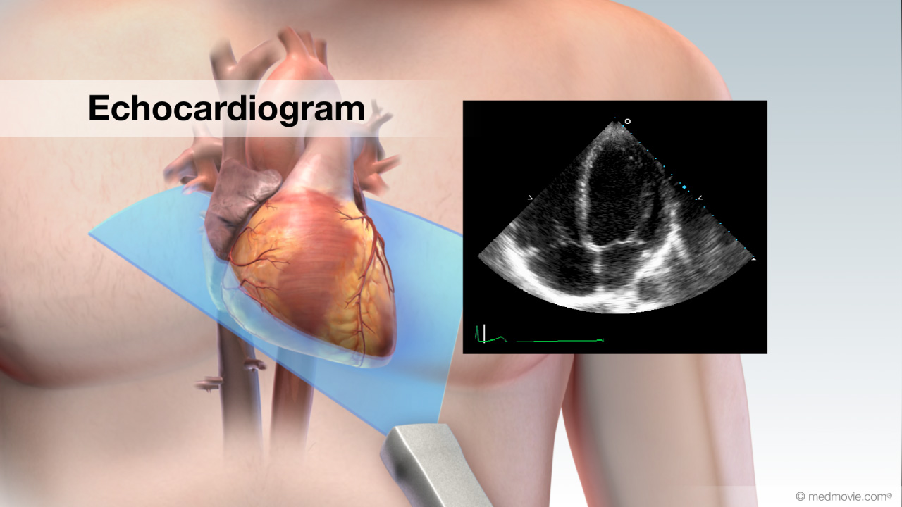
During an echo we use high-frequency sound waves to create computerized pictures of your heart and its attached blood vessels.

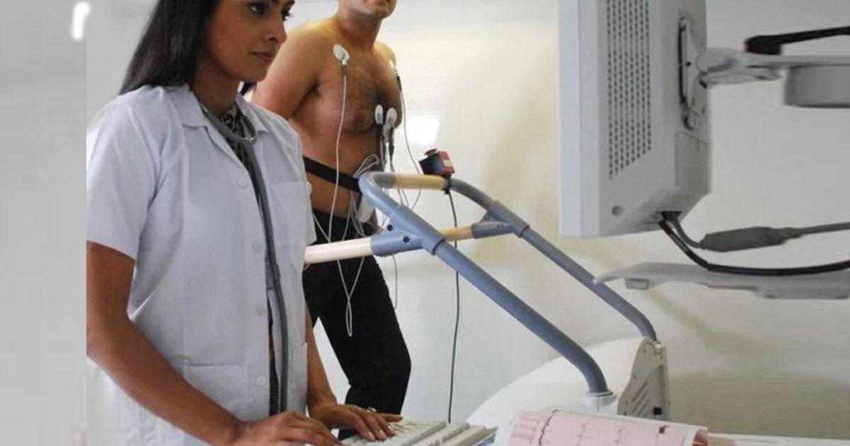
Heart test with ultrasound. It is most often used to detect a decrease in blood flow to the heart from narrowing in the coronary arteries. During an exercise stress test you are connected to a monitor and your blood pressure and heart rate are observed as you exercise on a treadmill. An echocardiogram or echo test is an ultrasound of your heart.
Echocardiograms are ultrasound tests which scan the heart and provide accurate information about the hearts structure and function. A heart ultrasound is a non-invasive way to get a look at how well the heart is working. It can be used on its own or in conjunction with an exercise stress test.
An echocardiogram echosound cardheart gramdrawing is an ultrasound test that can evaluate the structures of the heart as well as the direction of blood flow within it. These images help your doctor see your hearts chambers valves and. Ultrasound uses sound waves beyond the range of human hearing to image the chambers and walls of the heart.
Stress echocardiography is a test that uses ultrasound imaging to show how well your heart muscle is working to pump blood to your body. An echocardiogram echo is a graphic outline of the hearts movement. Home Diagnostic Testing Ultrasounds Dopplers.
It can help your healthcare provider see how well your lungs and heart are working. During an echocardiogram a technician uses a probe that emits high-frequency sound waves ultrasound that echo off the structures of your heart. Ultrasounds and dopplers are diagnostic tests used to analyze the overall function of your heart and to detect the presence of many different types of heart disease.
The waves which are translated into video images visible on a monitor can reveal in-formation about your hearts structure and function. How the Test is Performed This test is done at a medical center or health care providers office. Echo is often combined with Doppler ultrasound and color Doppler to evaluate blood flow across the hearts.
How is a heart ultrasound done. An echocardiogram echo is a test that uses high frequency sound waves ultrasound to make pictures of your heart. During an echo test ultrasound high-frequency sound waves from a hand-held wand placed on your chest provides pictures of the hearts valves and chambers and helps the sonographer evaluate the pumping action of the heart.
Images of the heart are taken during a stress echocardiogram to see if. A stress echocardiogram tests how well your heart and blood vessels are working especially under stress. The test is also called echocardiography or diagnostic cardiac ultrasound.
A probe is used to send sound waves from. In some cases physicians recommend going directly to a Thallium Exercise Stress Test and bypassing the Echo Exercise Stress Test. An echocardiogram is a test that uses ultrasound to show how your heart muscle and valves are working.
A chest ultrasound is an imaging test that uses sound waves to look at the structures and organs in your chest. The test is attempting to understand how the heart is working while resting.


